Introduction
With the generous support of Dumbarton Oaks, Byzantine Studies Program, Drs. Ioanna Kakoulli and Christian Fischer, professors in the Department of Materials Science and Engineering at the University of California Los Angeles, with joint appointment in the UCLA/Getty Interdepartmental Degree Program on the Conservation of Archaeological and Ethnographic Materials, were able to complete the second phase of in situ analysis of the important and unique wall paintings at the Enkleistra of St. Neophytos in Cyprus. This campaign involved the reassessment of ambiguous results (using scientific imaging and portable spectroscopic instruments) and selective sampling from representative areas of the paintings for micro-analysis. Samples were prepared as polished cross-sections in the laboratory and examined using polarized light microscopy (PLM) as well as scanning electron microscopy (SEM) at variable pressure (VP) coupled with energy dispersive x-ray spectroscopy (EDS). Selected samples were also analyzed using Fourier transform infrared spectroscopy (FTIR). Owing to the complex nature of the paintings, the ambiguous dating of the different painting phases and the varied and convoluted techniques, the focus of this campaign was to complete the scientific measurements and documentation that would help determine unresolved matters. This project builds upon previous research (conducted between 2008 and 2010) supported by Dumbarton Oaks and the UCLA Senate and was particularly successful at answering questions raised during previous research and also leading to new discoveries.See here for the report for the 2008/09 campaign. I. Kakoulli and C. Fischer, in Enkleistriotika Analekta. Proceedings of the 1st International Conference St. Neophytos Enkleistros, History-Theology-Civilization, edited by A. Archimandrite Enkleistriotis, 437–61 (Paphos: Ieras Vasilikis kai Stavropigiakis monis Agiou Neophytou, 2011).
Brief Overview of the Enkleistra and the Wall Paintings
")
")
Renowned for the wall paintings adorning the chambers carved into the limestone cliffs, the Enkleistra ("place of reclusion") “preserves the personal stamp of its founder” in the representation of the saint in the wall painting iconography and provides a “rare exception in the vast impersonality of Byzantine art." In 1183, Theodore Apseudes executed the wall paintings in the saint's cell and the Bema in the “classic court style” based on the "rococo" manner of painting, which appears to have been evolved in Constantinople in the last quarter of the twelfth century. The attribution of these paintings to Theodore Apseudes was supported by an inscription written on the north wall of the cell that reads: “the Enkleistra was painted by the hand of me Theodore Apseudes, in the year 6691 from Adam [1183].” Based on written testimonies, the walls of the Naos were painted after 1197 in the “monastic style,” in which the figures are drawn with austere and hard lines, by an unknown painter in the “Comnenian provincial” or the “linear style."C. A. Mango and E. J. W. Hawkins, "The Hermitage of St. Neophytos and Its Wall Paintings," Dumbarton Oaks Papers 20 (1966): 119–206, especially 121–22, 129, 183, 194, and 198.
Our previous research based solely on non-invasive techniques indicated the presence of numerous painting phases (not always easy to discern) with a terminus post quem of 1160, marking the completion of the excavation of the Cell, and a terminus ante quem of the middle of the eighteenth century, corresponding to the opening of the door in the east wall of the Cell.See above, n. 1, and I. Kakoulli, C. Fischer, D. Michaelides, in IIC, Istanbul Congress, edited by C. Rozeik, A. Roy, and D. Saunders, 1:96–102 (Istanbul: The International Institute for Conservation of Historic and Artistic Works, 2010). Within this time frame, there are several overlaying painting phases, not all of which have been previously recorded.
Wall Painting Techniques and Materials
")
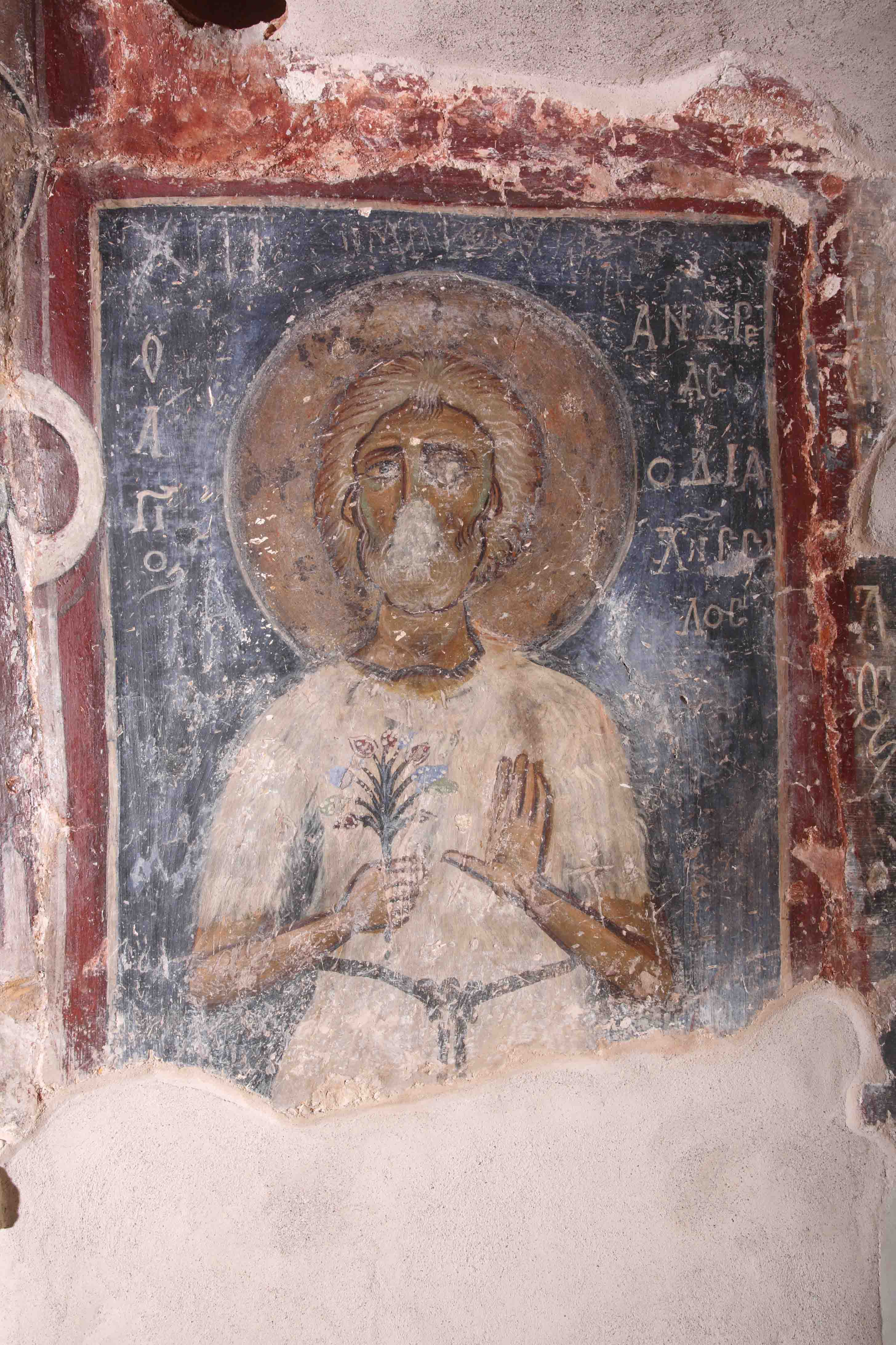
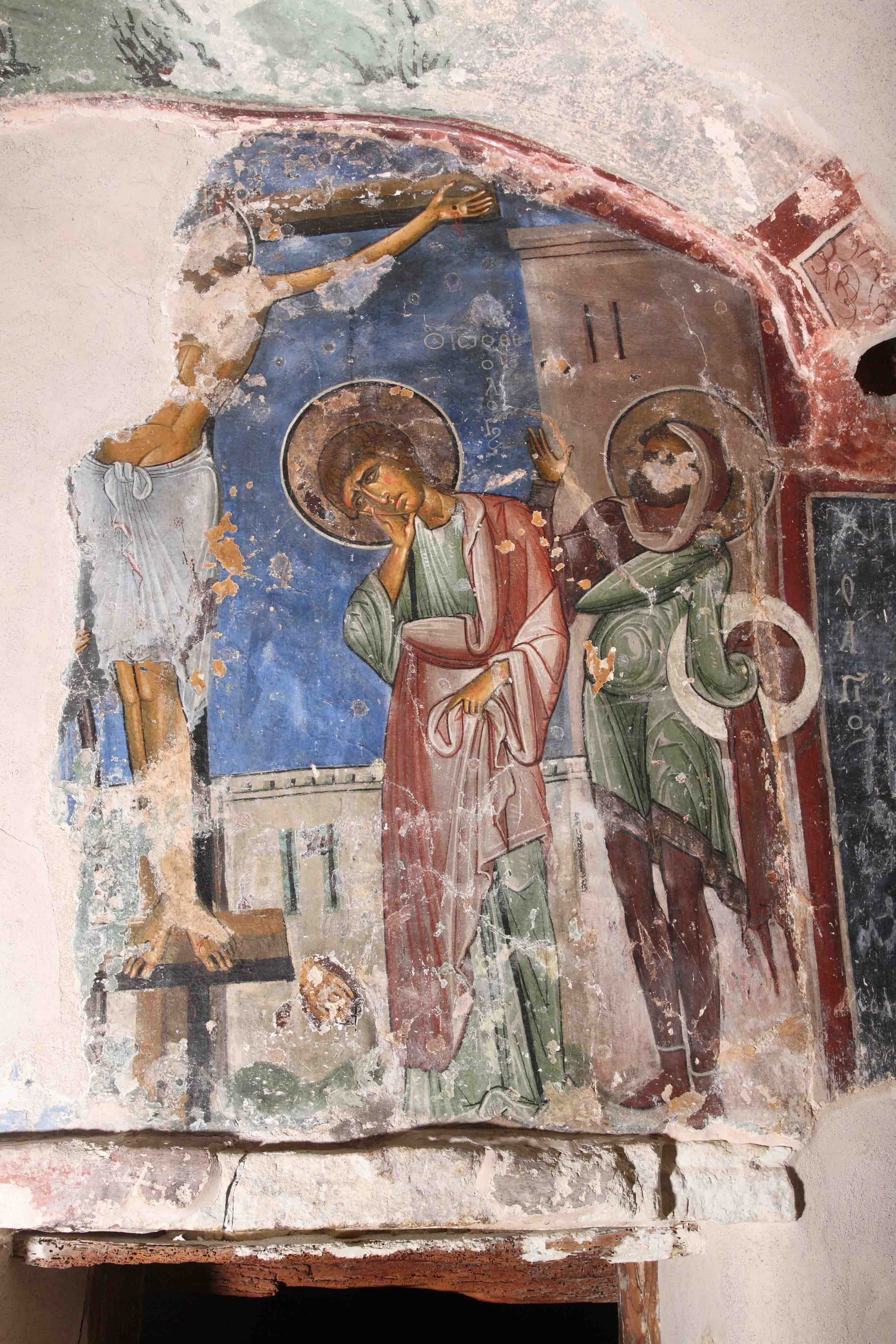
Two different techniques of the paintings were used in the Enkleistra: the fresco and the secco. The fresco technique can be clearly seen in the execution of the image of St. Andrew Salos painted on the south wall of the Cell. The panel was executed on an individual giornata of plaster with pigments compatible to the fresco technique. Conversely, the scene of the Crucifixion, painted on the same wall, was completed entirely in the secco technique. The painting was executed over a thin lead white layer characteristic to icon painting on wood applied over a preexisting painting. The pigments were mixed and applied on the wall using an organic binding medium, most likely a drying oil and/or egg tempera or glue. While an in-depth analysis of the chemical nature of the pigments and organic binding media employed in the painted decoration of the Enkleistra is in progress, data based primarily on noninvasive methods of analysis supported by nondestructive micro-analytical techniques have indicated the presence of various natural mineral pigments (local and foreign), as well as artificially produced inorganic pigments.
")
The local mineral varieties included haematite (Fe2O3) and goethite (iron (III) oxides and hydroxide - FeO(OH)), celadonite (a phyllosilicate of potassium, iron, aluminum and hydroxide - K(Mg,Fe2+)(Fe3+,Al)Si4O10(OH)2), calcium carbonate white (CaCO3), and possibly white clay.C. Klein and J. D. Dana, Manual of Mineralogy: (after James D. Dana) (New York, 1993). The foreign natural inorganic pigments included lapis lazuli ((Na,Ca)8(AlSiO4)6(S,SO4,Cl)1-2),J. Plesters, in Artists’ Pigments: A Handbook of Their History and Characteristics, edited by A. Roy, 2:37–61 (Washington, DC: National Gallery of Art, 1993). cinnabar (mercuric sulfide - HgS), and perhaps an arsenic-containing pigment, originally presumed to be orpiment (arsenic sulfide As2S3) or realgar (As4S4). The artificially-produced inorganic pigments on the other hand were lead white (basic lead carbonate - (PbCO3)2·Pb(OH)2) and minium or red lead (lead tetroxide - Pb3O4). The pigments seem to have been used pure or in mixtures and were applied in one or more layers. Thin metal foils of alloys containing gold and silver (electrum) were also identified in the paint layers and more particularly in the halos, where they added a metallic glitter.
Previous restorations and surface treatments using wax and modern polymers were also identified using noninvasive imaging and spectroscopy and confirmed by micro-Fourier transform infrared spectroscopy (µFTIR). The identification of the original pigments as well as the foreign substances on the surface of the paintings using non-contact probes reduced significantly the number of samples required for further study.
")
As a result, some 40 micro-samples were collected from representative areas of the entire extensive painted decoration of the Enkleistra for further analysis using non-destructive methods employing state-of-the-art instrumentation. Current investigations based on micro-analytical studies of both paint and plaster samples aim to identify the chemistry and provenance of pigments, the aggregates in the plasters, the layout techniques, and the treatment of the paints (mixtures of pigments, superposition of paint layers) in the various painting phases. These will shed new light on intra- and interregional exchange and raw-materials procurement, the techniques of application, and will also help identify markers characteristic to the different painting phases for more accurate attributions. Peculiarities and abnormalities in the initial results such as the presence of arsenic in a particular green paint found in the paintings both in the Cell and the Naos, as well as paint alterations and particular types of plaster aggregates, were further investigated through additional in situ measurements, sampling, and microanalysis.
Reassessment of Ambiguous Results from Previous Noninvasive Investigations
Investigations of the green paint containing arsenic
")
During XRF analysis of the pigments, some green areas painted with celadonite (green earth) as previously identified using UV/Vis/NIR spectroscopy indicated the prominent presence of arsenic in addition to the characteristic x-rays of Fe, a major element in the structure of celadonite. It is important to note that the green paint in other paintings attributed to the same painting phase, on stylistic grounds, do not contain arsenic.
The paintings with iron-rich green paints where so far arsenic was detected are:
- The green background in the Deesis scene on the north wall of the cell
- The green background in the scene of a standing Virgin Mary, orans, on the east wall of the Bema
- The green background in the crucifixion and deposition scenes on the east wall of the Naos.
While the presence of such a distinctive paint (implying some kind of correlation among the different phases of painting) is peculiar enough from an art historical perspective, the occurrence of arsenic in a green earth paint layer is also technologically unusual.
")
At first, the detection of arsenic (together with iron, calcium and small quantities of sulfur) in the green paint using portable x-ray fluorescence spectroscopy was connected to the possible presence of arsenopyrite (iron arsenic sulfide - FeAsS) or lollingite (iron arsenide - FeAs2) or to a deliberate admixture of green earth with realgar or orpiment (two relatively common minerals used as pigments) to change the hue of the green paint. To verify this hypothesis, a sample from this green paint was mounted as cross section and analyzed using VPSEM-EDS. The results from the analysis indicated that arsenic was associated with particles containing calcium but not iron. These arsenic/calcium-rich particles were dispersed in the green matrix but could not be differentiated under the PLM. VPSEM using the backscattered electron detector for compositional contrast and elemental mapping using EDS pointed towards the presence of an arsenic/calcium compound. This could indicate the presence of a calcium arsenate (pharmacolite?) mineral or another compound rich in arsenic and calcium. The results from the analysis were surprising and remain inconclusive; further analysis is currently in progress to identify the precise chemical composition and origin of this arsenic/calcium compound using Fourier Transform infrared and Raman spectroscopy, while all other samples with arsenic content are also investigated.
Composition of Plasters
In addition to the paint layers, the plaster layers as well have shown interesting initial results and compositions unusual to the Byzantine tradition of the late 12th century. During the study of the paintings at the Enkleistra, different samples were collected from plaster layers corresponding to different painting phases for comparison. Most of the plaster layers consist of calcium carbonate mixed with chopped straw and some sand, though in two painting phases—the painting depicting Christ on the north wall in the Naos (next to the iconostasis), attributed by Mango and Hawkins to the late 12th or the beginning of the 13th century, and the paintings in the Narthex (west wall)—have unusual compositions. The results from this analysis provide new information for the technology of Byzantine wall paintings from the late 12th century and early 13th century and will be communicated in a forthcoming publication.
Environmental Causes of Deterioration and Color Change
At the Enkleistra of St. Neophytos, the most imminent natural threats to the wall paintings are:
- The instability of the bedrock
- Human activity and an uncontrolled microclimate (RH, T, light, pollutants) due to the large number of tourists arriving at the site and entering the enclosed space of the tomb at the same time
- Seismic activity
The paintings suffer primarily from the movement of the bedrock, especially those in the Naos, which show alarming structural cracks and extensive loss of paint. Although religious services take place in the main church of the monastery, the fame of the Enkleistra draws large numbers of tourists and pilgrims. Extensive visitation has led to different degrees of physical damage to the wall paintings. This damage includes graffiti (dated to at least as early as the seventeenth century), scratching in the belief of miraculous healing powers attributed to the paint, defacing due to religious conflicts, religious practices, and fading and discolorations due to changes in the microclimate. In addition, micro-flaking and different types of paint alterations were observed on both the twelfth-century and post-Byzantine schemes. The most common alteration pattern documented was the darkening of the bright red paint layers containing red lead and cinnabar.
Discoloration and darkening of red paint layers containing lead pigments and cinnabar
")
It is widely accepted that the majority of the transformations and color changes in wall paintings are principally induced by environmental exposure factors such as light, humidity, pH, and microbial activity, assisted in some cases by the action of different deteriogens such as sodium chloride (NaCl), often present as a soluble salt, and deriving either from the constituent materials (wall, plaster, paint) as an impurity or as an external contaminant from the environment. Particularly, cinnabar/vermilion and lead-containing paint layers have been known to discolor to a dark gray/black color.K. Keune and J. J. Boon, AnalChem 77 (2006): 4742; M. Cotte et al., Talanta 70 (20060: 1136; Ibid., AnalChem 78 (2006): 7484; R. J. Gettens and G. L. Stout, Painting Materials: A Short Encycloaedia (New York, 1966); M. Cotte, E. Checroun, J. Susini, and P. Walter, Applied Physics A: Materials Science & Processing 89 (2007): 841; M. Spring and R. Grout, The National Gallery Technical Bulletin 23 (2002): 50; R. J. Gettens, R. L. Feller, and W. T. Chase, StudConserv 17 (1972): 45; S. Giovannoni, M. Matteini, and A. Moles, StudConserv 35 (1990): 21; R. Grout and A. Burnstock, Zeitschrift für Kunsttechnologie und Konservierung 141 (2000): 15; B. Pal, S. Ikeda, and B. Ohtani, Inorg Chem 42 (2003): 1518; M. Ravichandran, G. R. Aiken, M. M. Reddy, and J. N. Ryan, Environmental Science & Technology 32 (1998): 3305; S. Aze, J. M. Vallet, V. Detalle, O. Grauby, and A. Baronnet, Phase Transitions 81 (2008): 145; S. B. Aze, J.-M. Vallet, A. Baronnet, and O. Grauby, Eur J Mineral 18 (Nov/Dec 2006): 835, 883.
At the Enkleistra of St. Neophytos, alterations of red paints were observed macroscopically as discolored zones and microscopically on stratigraphic samples as black and hazy particles, forming in some cases distinct layers. These observations agree with published literature on the HgS and Pb3O4 altering from red into grey/black. Five samples, all containing red lead (Pb3O4) and/or cinnabar (HgS), and with evident color change, were taken for analysis from different painting phases.Samples taken: S08 (Naos, red book, figure of Christ, north wall, eastern side); S16 (Cell, north wall niche, roundel with cross); S29 (Bema, dome with Pantokrator, altered red from the border of the roundel with the Virgin); S30 (Bema, dome with Pantokrator, altered red from the mandorla); and S40 (Naos, west wall, 1503, washing of the feet scene, red altered garment). The samples were prepared as polished cross sections and analyzed using PLM and VPSEM-EDS.
 (Koukoulli and Fischer 2011–2012)")
Alteration of cinnabar into meta-cinnabar has been noted as a change in the crystal structure from hexagonal to cubic or amorphous state.I. Istudor, A. Dină, G. Rosu, D. Seclăman, and G. Niculescu, in e-conservation magazine (2004). Upon exposure to halide solutions, cinnabar has been documented to undergo photochemical darkening.J. K. McCormack, Mineralium Deposita 35 (2000): 796. At the Enkleistra, the secluded environment and its potential for seepage of water make this a possible mechanism for the transformation of cinnabar. However, the exact composition and phase of the altered material remains to be identified.
Lead red undergoes a transformation from red to black like cinnabar. Previous studies through XRD on this alteration found that lead red Pb3O4 transforms into plattnerite (PbO2) and also anglesite (PbSO4).S. Aze, J.-M. Vallet, M. Pomey, A. Baronnet, and O. Grauby, European Journal of Mineralogy 19 (2007): 883. It has been suspected that acidic environments and SO2 pollutants, a precursor to acid rain, can lead to the solvation of the Pb and to the formation of the altered products. Given the prevalence of these pollutants at the Enkleistra, this is a possible mechanism for this alteration of the analyzed samples. Likewise, as these are common elements phase analysis is necessary to determine whether the plattnerite and anglesite are the altered products.
Conclusion
The paintings of St. Neophytos present a microcosm of Byzantine painting traditions and a complex ecosystem. It is therefore important to study the paintings not only in their art historical context but also in their architectural and natural environment. The analysis of the paintings is a meticulous and slow operation. The next phases of this project will focus on completing the analysis of all samples in the laboratory, completing the documentation, and subsequently imaging the different painting phases with terahertz technology to see behind the top plaster layers, something that is impossible with infrared or other radiation.
Acknowledgements
We would like to express our gratitude to Dumbarton Oaks for supporting the study of these important paintings at the Enkleistra of St. Neophytos; to St. Neophytos Monastery, the abbot, the monks, and more particularly to Archimandrite Alexios Enkleistriotis for their kind permission to study the paintings and warm hospitality; and to the Department of Antiquities of Cyprus for the permission to carry out the scientific investigation of the paintings.
-
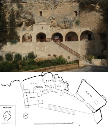
 Fig. 1: Exterior of the Enkleistra and plan of the cave complex (Koukoulli and Fischer 2011–2012)
Fig. 1: Exterior of the Enkleistra (top) and plan of the cave complex showing the Cell, the Bema, the Naos, and the Narthex (bottom; Mango and Hawkins 1966).
Fig. 1: Exterior of the Enkleistra and plan of the cave complex (Koukoulli and Fischer 2011–2012)
Fig. 1: Exterior of the Enkleistra (top) and plan of the cave complex showing the Cell, the Bema, the Naos, and the Narthex (bottom; Mango and Hawkins 1966).
-
 Fig. 1a: Plan of the Enkleistra complex of St. Neophytos (Koukoulli and Fischer 2011–2012)
Fig. 1a: Plan of the Enkleistra complex of St. Neophytos (Mango and Hawkins 1966).
Fig. 1a: Plan of the Enkleistra complex of St. Neophytos (Koukoulli and Fischer 2011–2012)
Fig. 1a: Plan of the Enkleistra complex of St. Neophytos (Mango and Hawkins 1966).
-
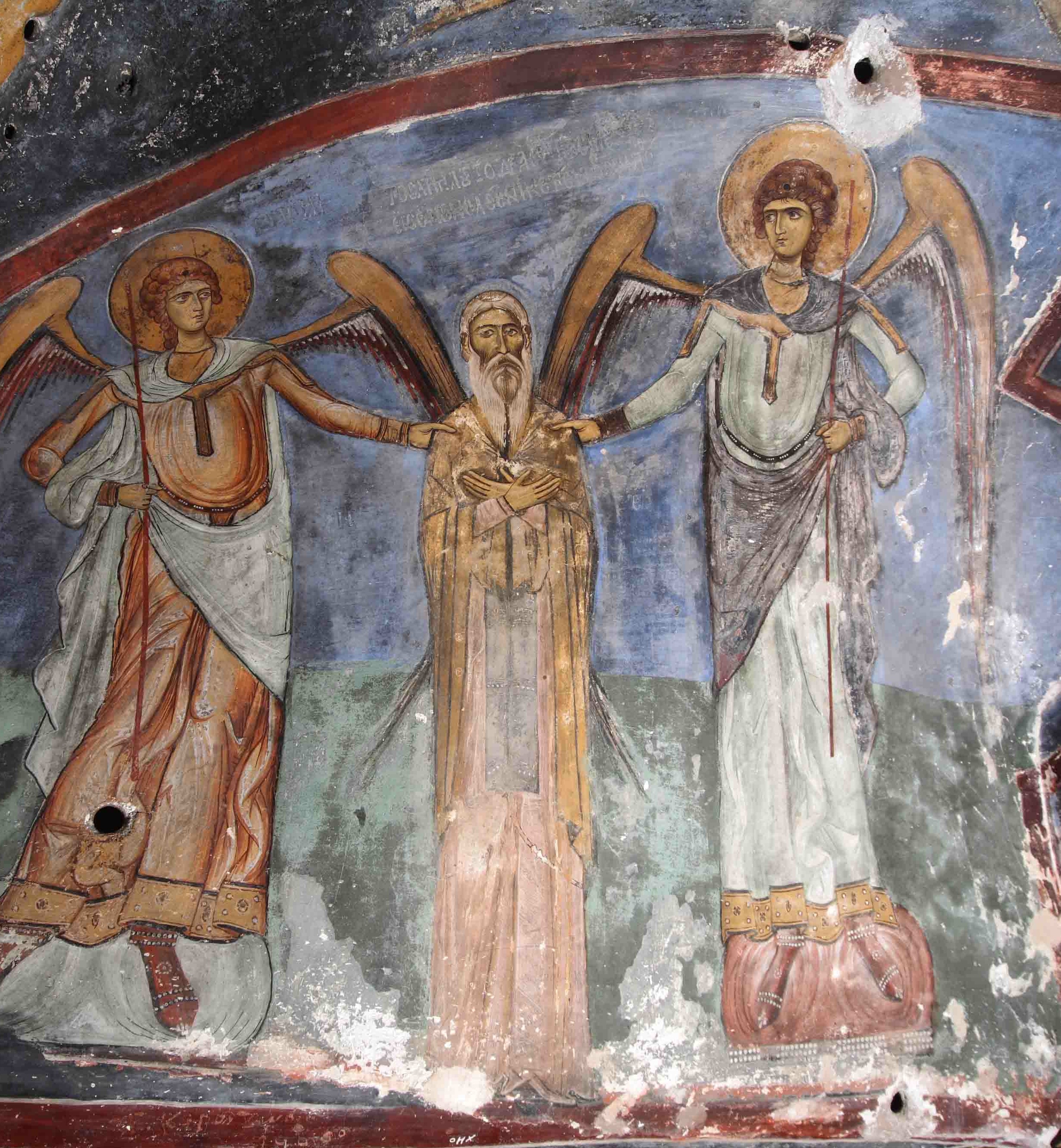
 Fig. 2: Depiction of St. Neophytos flaked by the archangels Michael and Gabriel (Koukoulli and Fischer 2011–2012)
Fig. 2: Depiction of St. Neophytos flaked by the archangels Michael and Gabriel, who hold him by the shoulders (Bema).
Fig. 2: Depiction of St. Neophytos flaked by the archangels Michael and Gabriel (Koukoulli and Fischer 2011–2012)
Fig. 2: Depiction of St. Neophytos flaked by the archangels Michael and Gabriel, who hold him by the shoulders (Bema).
-
 Fig. 3: Fresco painting depicting St. Andrew Salos (Koukoulli and Fischer 2011–2012)
Fig. 3: Fresco painting depicting St. Andrew Salos, painted in a single giornata.
Fig. 3: Fresco painting depicting St. Andrew Salos (Koukoulli and Fischer 2011–2012)
Fig. 3: Fresco painting depicting St. Andrew Salos, painted in a single giornata.
-
 Fig. 4: Scene of the Crucifixion on the south wall of the Cell (Koukoulli and Fischer 2011–2012)
Fig. 4: Scene of the Crucifixion on the south wall of the Cell, painted over a thin layer of lead white applied as an intermediate layer to mask a preexisting painting.
Fig. 4: Scene of the Crucifixion on the south wall of the Cell (Koukoulli and Fischer 2011–2012)
Fig. 4: Scene of the Crucifixion on the south wall of the Cell, painted over a thin layer of lead white applied as an intermediate layer to mask a preexisting painting.
-
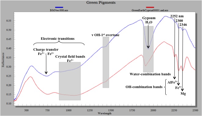
 Fig. 5: UV/Vis/NIR reflectance spectra of a reference celadonite mineral sample from Cyprus and an unknown green paint sample from the Enkleistra (Koukoulli and Fischer 2011–2012)
Fig. 5: UV/Vis/NIR reflectance spectra of a reference celadonite mineral sample from Cyprus and an unknown green paint sample from the Enkleistra. UV/Vis/NIR provided the exact identification of celadonite with the speciation of the iron (Fe) compounds showing a broad absorption between 540 and 750 nm indicative of the d-d ligand-field and intervalence charge-transfer (IVCT) transitions in the visible region of the chromophors Fe^+2 and Fe^+3 and by the overtones and combination bands of the structural OH (octahedral site, bound to a cation) stretching and bending vibrations and H2O stretching and bending vibrations in the near infrared region.
Fig. 5: UV/Vis/NIR reflectance spectra of a reference celadonite mineral sample from Cyprus and an unknown green paint sample from the Enkleistra (Koukoulli and Fischer 2011–2012)
Fig. 5: UV/Vis/NIR reflectance spectra of a reference celadonite mineral sample from Cyprus and an unknown green paint sample from the Enkleistra. UV/Vis/NIR provided the exact identification of celadonite with the speciation of the iron (Fe) compounds showing a broad absorption between 540 and 750 nm indicative of the d-d ligand-field and intervalence charge-transfer (IVCT) transitions in the visible region of the chromophors Fe^+2 and Fe^+3 and by the overtones and combination bands of the structural OH (octahedral site, bound to a cation) stretching and bending vibrations and H2O stretching and bending vibrations in the near infrared region.
-
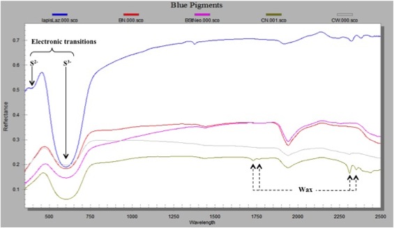
 Fig. 6: UV/Vis/NIR reflectance spectra of a reference lapis lazuli (lazurite mineral) and unknown blue paint samples from the Enkleistra (Koukoulli and Fischer 2011–2012)
Fig. 6: UV/Vis/NIR reflectance spectra of a reference lapis lazuli (lazurite mineral) and unknown blue paint samples from the Enkleistra. The identification of lapis lazuli in the blue paints was determined by its distinctive electronic spectrum displaying a strong and broad absorption around 600 nm in the visible caused by the S3 paramagnetic radical anion in lapis lazuli.
Fig. 6: UV/Vis/NIR reflectance spectra of a reference lapis lazuli (lazurite mineral) and unknown blue paint samples from the Enkleistra (Koukoulli and Fischer 2011–2012)
Fig. 6: UV/Vis/NIR reflectance spectra of a reference lapis lazuli (lazurite mineral) and unknown blue paint samples from the Enkleistra. The identification of lapis lazuli in the blue paints was determined by its distinctive electronic spectrum displaying a strong and broad absorption around 600 nm in the visible caused by the S3 paramagnetic radical anion in lapis lazuli.
-
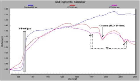
 Fig. 7: UV/Vis/NIR reflectance spectra of a reference cinnabar sample and unknown red paint samples from the Enkleistra (Koukoulli and Fischer 2011–2012)
Fig. 7: UV/Vis/NIR reflectance spectra of a reference cinnabar sample and unknown red paint samples from the Enkleistra. The identification of cinnabar in the red paints was possible due to the characteristic absorption edge (or band edge) of sulfur (S) and by pXRF spectroscopy with the photon emissions at 9.987 keV (L-alpha edge energy) and 11.823 keV (L-beta edge energy) characteristic of mercury.
Fig. 7: UV/Vis/NIR reflectance spectra of a reference cinnabar sample and unknown red paint samples from the Enkleistra (Koukoulli and Fischer 2011–2012)
Fig. 7: UV/Vis/NIR reflectance spectra of a reference cinnabar sample and unknown red paint samples from the Enkleistra. The identification of cinnabar in the red paints was possible due to the characteristic absorption edge (or band edge) of sulfur (S) and by pXRF spectroscopy with the photon emissions at 9.987 keV (L-alpha edge energy) and 11.823 keV (L-beta edge energy) characteristic of mercury.
-
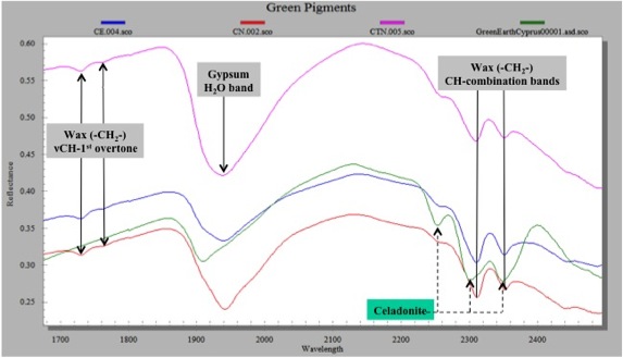
 Fig. 8: UV/Vis/NIR reflectance spectra of a green paint samples that were covered by wax applied in a previous restoration (Koukoulli and Fischer 2011–2012)
Fig. 8: UV/Vis/NIR reflectance spectra of a green paint samples that were covered by wax applied in a previous restoration. The wax was identified through the characteristic absorptions of CH2 and CH combination bands and overtones in the near infrared.
Fig. 8: UV/Vis/NIR reflectance spectra of a green paint samples that were covered by wax applied in a previous restoration (Koukoulli and Fischer 2011–2012)
Fig. 8: UV/Vis/NIR reflectance spectra of a green paint samples that were covered by wax applied in a previous restoration. The wax was identified through the characteristic absorptions of CH2 and CH combination bands and overtones in the near infrared.
-
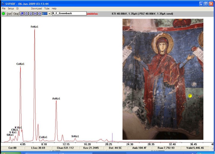
 Fig. 9: XRF spectrum of the green paint in the scene depicting Virgin Mary (Koukoulli and Fischer 2011–2012)
Fig. 9: XRF spectrum of the green paint in the scene depicting Virgin Mary, orans (Bema, east wall), showing the prominent presence of arsenic (As). The iron (Fe) is from celadonite and calcium (Ca) from the plaster (calcium carbonate).
Fig. 9: XRF spectrum of the green paint in the scene depicting Virgin Mary (Koukoulli and Fischer 2011–2012)
Fig. 9: XRF spectrum of the green paint in the scene depicting Virgin Mary, orans (Bema, east wall), showing the prominent presence of arsenic (As). The iron (Fe) is from celadonite and calcium (Ca) from the plaster (calcium carbonate).
-
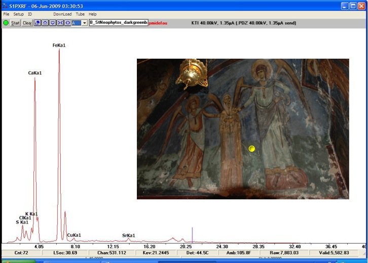
 Fig. 10: XRF spectra of the green paint in a scene in the Bema (Koukoulli and Fischer 2011–2012)
Fig. 10: XRF spectra of the green paint in a scene in the Bema that does not contain arsenic (As). The spectrum shows the characteristic x-rays for Fe in the celadonite and Ca in the plaster.
Fig. 10: XRF spectra of the green paint in a scene in the Bema (Koukoulli and Fischer 2011–2012)
Fig. 10: XRF spectra of the green paint in a scene in the Bema that does not contain arsenic (As). The spectrum shows the characteristic x-rays for Fe in the celadonite and Ca in the plaster.
-
 Fig. 11: XRF spectra of the green paint in a scene in the Bema (Koukoulli and Fischer 2011–2012)
Fig. 11: XRF spectra of the green paint in a scene in the Bema that does not contain arsenic (As). The spectrum shows the characteristic x-rays for Fe in the celadonite and Ca in the plaster.
Fig. 11: XRF spectra of the green paint in a scene in the Bema (Koukoulli and Fischer 2011–2012)
Fig. 11: XRF spectra of the green paint in a scene in the Bema that does not contain arsenic (As). The spectrum shows the characteristic x-rays for Fe in the celadonite and Ca in the plaster.
-
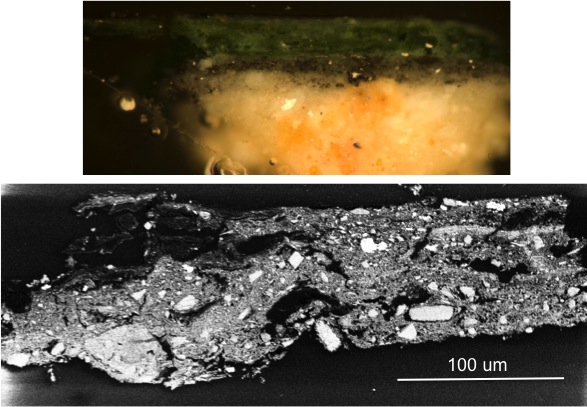
 Fig. 12: Cross section samples of the arsenic-rich green paint (Koukoulli and Fischer 2011–2012)
Fig. 12: Cross section samples of the arsenic-rich green paint (from the Deesis scene, Cell, north wall) in reflected polarized light (top) and in backscattered electrons using VPSEM (bottom).
Fig. 12: Cross section samples of the arsenic-rich green paint (Koukoulli and Fischer 2011–2012)
Fig. 12: Cross section samples of the arsenic-rich green paint (from the Deesis scene, Cell, north wall) in reflected polarized light (top) and in backscattered electrons using VPSEM (bottom).
-
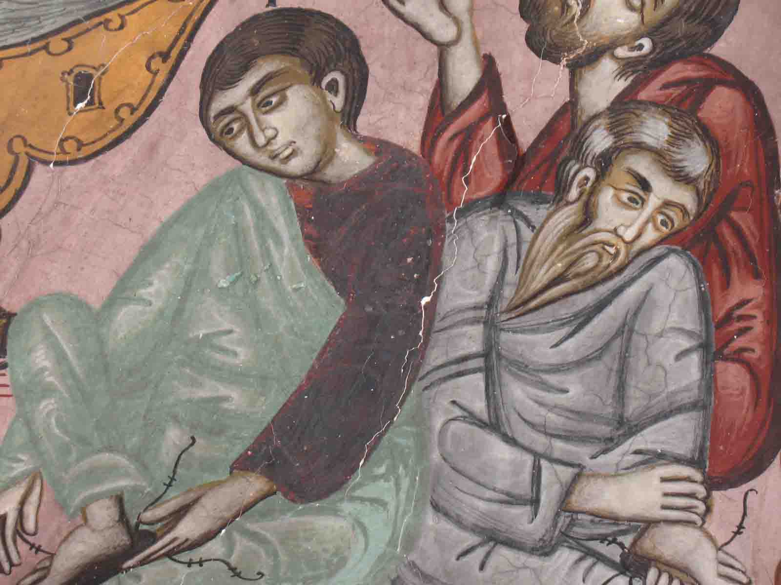
 Fig. 13: Image of the scene “washing of the feet” (Koukoulli and Fischer 2011–2012)
Fig. 13: Image of the scene “washing of the feet” (Naos, west wall), showing an extensive area of red alteration on the robe of the apostle to the left.
Fig. 13: Image of the scene “washing of the feet” (Koukoulli and Fischer 2011–2012)
Fig. 13: Image of the scene “washing of the feet” (Naos, west wall), showing an extensive area of red alteration on the robe of the apostle to the left.
-
 Fig. 14: Alteration of cinnabar (HgS) (Koukoulli and Fischer 2011–2012)
Fig. 14: Alteration of cinnabar (HgS). Micrographs of sample 16 (from left clockwise): a stereomicroscopic image of sample 16 showing a layer of altered red; the same sample.
Fig. 14: Alteration of cinnabar (HgS) (Koukoulli and Fischer 2011–2012)
Fig. 14: Alteration of cinnabar (HgS). Micrographs of sample 16 (from left clockwise): a stereomicroscopic image of sample 16 showing a layer of altered red; the same sample.